
Michelle Mendoza
Associate Professor of Oncological Sciences and Adjunct Associate Professor of Biomedical Engineering
Cell Migration, Invasion, RAS, ERK, Lung Cancer, Signaling, Tumor Mechanics

Molecular Biology Program
Education
B.S. Pennsylvania State University
Ph.D. University of California, San Diego
Research
The Mendoza lab mission is to help patients with lung cancer and other solid tumors through the discovery of fundamental mechanisms of cancer progression. We apply physical science perspectives to understand biochemical and biomechanical signaling in tumor cells and their microenvironment.
We are interested in the signaling of cell migration and cancer invasion. Questions addressed in the lab include:
- How do signaling pathways synergistically control actin and adhesion dynamics for cell movement?
- How biomechanical forces cooperate with ERK signaling to drive lung cancer invasion?
- How do activated fibroblasts communicate with tumor cells to promote cancer progression?
We approach these and other biological problems with a combination of experimental
and computational techniques, including biochemistry, quantitative imaging, simulations,
and mouse models. Students in the lab receive interdisciplinary training spanning
cancer biology, biomechanics, and translational science through collaborations across
Huntsman Cancer Institute and the University of Utah
Cell migration requires coordinated regulation of actin assembly, adhesion dynamics,
and force generation. We investigate how signaling pathways integrate these processes
to direct cell movement, and how this is activated during cancer invasion.
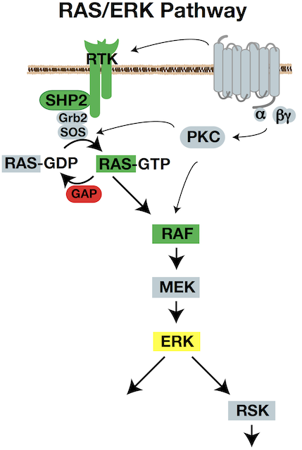
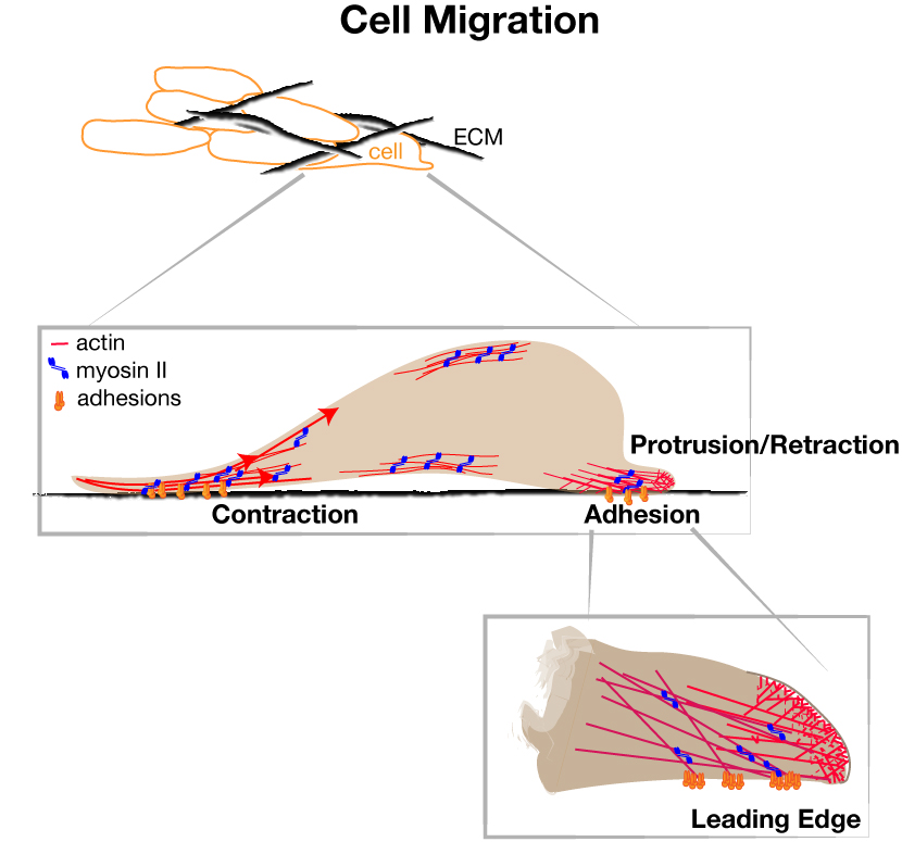
Integrated Forces for Cell Migration
How do cells convert signaling information into physical movement? Our laboratory
investigates the molecular and mechanical mechanisms that power cell migration. We
have shown that ERK signaling coordinates actin assembly, adhesion dynamics, and cellular
contractility to generate productive movement. Current projects explore how these
forces are integrated across cells and their surrounding extracellular matrix, providing
new insight into both normal cell behavior and the invasive migration of cancer cells.
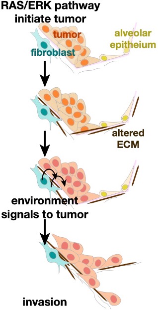
Tumor Biomechanics and Cancer Invasion
When cancers spread throughout the body, tumor cells navigate altered tumor microenvironments
and foreign structures. A computational modeling collaboration with the Weiss lab
in Biomedical Engineering showed that tensile strain can be amplified at the edge
of early lung tumors and in the tumor microenvironment. We are characterizing the
roles of strain signaling in early lung tumors by experimentally stretching tumor
cells and tumor-associated fibroblasts. With continued modeling, we are identifying
mechanisms by which the altered mechanical forces promote tumor invasion.
Lung Cancer Progression
Our lung cancer research also seeks to understand how environmental exposures interact
with genetic mutations to drive disease progression. We study how toxic exposures
and tissue injury promote fibroblast activation, extracellular matrix remodeling,
and tumor progression. Students in the lab use a combination of mouse models, human
tissue, quantitative imaging, and collaborations with population scientists to understand
how these interactions cause aggressive lung cancer phenotypes and resistance to therapeutics.
References
- ERK builds a population of short-lived nascent adhesions that produce persistent edge protrusion and cell migration. Andrew P. Shepherd, Keith R. Carney, Majid Rostami, Jakaria Shawon, Andrew Elliott, Shiela C. Samson, Sangyoon J. Han, and Michelle C. Mendoza. (2026) Proc Natl Acad Sci U S A. 123(25):e2525452123. https://www.pnas.org/doi/10.1073/pnas.2525452123
- The LOK C-terminus is an IBAR-like domain that facilitates membrane binding and ezrin colocalization. Jeriann R. Beiter Beiter, Dovydos Vasiliauskas, Gillian M. Hodge, Hannah E. Schneiderman, Monica C. Pillon, Sahithya S. Iyer, Michelle C. Mendoza, Andrew T. Lombardo, and Gregory A. Voth. (2026) Biophys J. 125(11):2541-2554. https://pubmed.ncbi.nlm.nih.gov/41958020/
- On the Mechanism of Ezrin Activation. Dovydos Vasiliauskas, Jeriann Beiter, Sahithya S. Iyer, Anthony T. Lombardo, Michelle C. Mendoza, and Gregory A. Voth. (2026) Biophys J. 18:S0006-3495(26)00216-X. https://pubmed.ncbi.nlm.nih.gov/41852029/
- Mechanism of ERK-mediated Rho Activation and Stress Fiber Assembly for Cell Migration. Akib M. Khan, Jakaria Shawon, Jared P. Bergman, Keith R. Carney, and Michelle C. Mendoza. (2025) BioRxiv. doi: 2025.11.15.688645. https://www.biorxiv.org/content/10.1101/2025.11.15.688645v1
- Tenascin-C in the early lung cancer tumor microenvironment promotes progression through integrin αvβ1 and FAK. Shiela C. Samson, Anthony Rojas, Rebecca G. Zitnay, Keith R. Carney, Wakeiyo Hettinga, Mary C. Shaelling, Delphine Sicard, Wei Zhang, Melissa Gilbert-Ross, Grace K. Dy, Michael J. Cavnar, Muhammad Furquan, Robert F. Browning Jr., Abdul R. Naqash, Bryan P. Schneider, Ahmad Tarhini, Daniel J. Tschumperlin, Alessandro Venosa, Adam I. Marcus, Lyska L. Emerson, Benjamin T. Spike, Beatrice S. Knudsen, and Michelle C. Mendoza. (2024) BioRxiv. doi: 2024.09.17.613509. https://www.biorxiv.org/content/10.1101/2024.09.17.613509v1
- Nonthreaded Isomers of Sungsanpin and Ulleungdin Lasso Peptides Inhibit H1299 Cancer Cell Migration. Lori Digal, Shiela C. Samson, Mark A. Stevens, Abhijit Ghorai, Hyungyu Kim, Marcus C. Mifflin, Keith R. Carney, David L. Williamson, Soohyun Um, Gabe Nagy, Dong-Chan Oh, Michelle C.Mendoza, and Andrew G. Roberts. (2024) ACS Chem Biol. 19(1):81-88. https://pubmed.ncbi.nlm.nih.gov/38109560/
- Nascent adhesions shorten the period of lamellipodium protrusion through the Brownian ratchet mechanism. Keith R. Carney, Akib M. Khan, Samantha Stam, Shiela C. Samson, Nikhil Mittal, Sangyoon J. Han, Tamara C. Bidone, and Michelle C. Mendoza.(2023) Mol Biol Cell. 34(12):ar115. https://pubmed.ncbi.nlm.nih.gov/37672339/
- Computational model of integrin adhesion elongation under an actin fiber. Samuel Campbell, Michelle C. Mendoza, Aravind Rammohan, Matthew E. McKenzie, and Tamara C. Bidone. (2023) PLoS Comput Biol. 19(7):e1011237. https://pubmed.ncbi.nlm.nih.gov/37410718/
- NKX2-1 controls lung cancer progression by inducing DUSP6 to dampen ERK activity.Kelley Ingram, Shiela C. Samson, Rediet Zewdu, Rebecca G. Zitnay, Eric L. Snyder, and Michelle C. Mendoza. (2022) Oncogene. 41(2):293-300. https://pubmed.ncbi.nlm.nih.gov/34689179/
- Mechanics of lung cancer: A finite element model shows strain amplification during early tumorigenesis. Rebecca G. Zitnay, Michael R. Herron, Keith R. Carney, Scott Potter, Lyska L. Emerson, Jeffrey A. Weiss, and Michelle C. Mendoza. (2022) PLoS Comput Biol. 18(10):e1010153. https://pubmed.ncbi.nlm.nih.gov/36279309/
- ERK signaling for cell migration and invasion. Shiela C. Samson, Akib, M. Khan, and Michelle C. Mendoza. (2022). Front. Mol. Biosci (9)998475. https://www.frontiersin.org/journals/molecular-biosciences/articles/10.3389/fmolb.2022.998475/full
- An NKX2-1/ERK/WNT feedback loop modulates gastric identity and response to targeted therapy in lung adenocarcinoma. Rediet Zewdu, Elnaz M. Mehrabad, Kelley Ingram, Pengshu Fang, Katherine L. Gillis, Soledad A. Camolotto, Grace Orstad, Alex Jones, Michelle C. Mendoza, Benjamin T. Spike, and Eric L. Snyder. (2021) Elife. 10:e66788. https://pubmed.ncbi.nlm.nih.gov/33821796/
- p90 ribosomal S6 kinase (RSK) phosphorylates myosin phosphatase and thereby controls edge dynamics during cell migration. Shiela C. Samson, Andrew Elliott, Brian D. Mueller, Yung Kim, Keith R. Carney, Jared P. Bergman, John Blenis, and Michelle C. Mendoza. (2019) Journal of Biological Chemistry 294(28):10846-10862. https://pubmed.ncbi.nlm.nih.gov/31138649/
- AKT1E17K Activates Focal Adhesion Kinase and Promotes Melanoma Brain Metastasis.David A. Kircher, Kirby A. Trombetti, Mark R. Silvis, Gennie L. Parkman, Grant M. Fischer, Stephanie N. Angel, Christopher M. Stehn, Sean C. Strain, Allie H. Grossmann, Keith L. Duffy, Kenneth M. Boucher, Martin McMahon, Michael A. Davies, Michelle C. Mendoza, Matthew W. VanBrocklin and Sheri L. Holmen. (2019) Mol Cancer Res. 9:1787-1800. https://pubmed.ncbi.nlm.nih.gov/31138602/
- ERK Reinforces Actin Polymerization to Power Persistent Edge Protrusion during Motility. Michelle C. Mendoza, Marco Vilela, Jesus E. Juarez, John Blenis, and Gaudenz Danuser (2015) Science Signaling 8:ra47. pubmed.ncbi.nlm.nih.gov/25990957/
- Phosphoregulation of the WAVE Regulatory Complex and Signal Integration. Michelle C. Mendoza. (2013) Seminars in Cell and Developmental Biology 24: 272-9. https://pubmed.ncbi.nlm.nih.gov/23354023/
- AKT Facilitates EGFR Trafficking and Degradation by Phosphorylating and Activating PIKfyve. E. Emrah Er, Michelle C. Mendoza, Ashley M. Mackey, Lucia E. Rameh, and John Blenis. (2013) Science Signaling 6:ra34. https://pubmed.ncbi.nlm.nih.gov/23757022/
- Quantitative Fluorescent Speckle Microscopy (QFSM) to Measure Actin Dynamics. Michelle C. Mendoza, Sebastien Besson, and Gaudenz Danuser. (2012) Current Protocols in Cytometry 62:2.18.1-2.18.25. https://pubmed.ncbi.nlm.nih.gov/23042526/
- ERK-MAPK Drives Lamellipodia Protrusion by Activating the WAVE2 Regulatory Complex. Michelle C. Mendoza, E. Emrah Er, Wenjuan Zhang, Bryan A. Baliff, Hunter L. Elliott, Gaudenz Danuser, and John Blenis. (2011) Molecular Cell 41:661-71. https://pubmed.ncbi.nlm.nih.gov/21419341/
- The Ras-ERK and PI3K-mTOR Pathways: Cross-talk and Compensation. Michelle C. Mendoza, E. Emrah Er, and John Blenis. (2011) Trends in Biomedical Sciences 36:320-8. https://pubmed.ncbi.nlm.nih.gov/21531565/
- ERK-MAPK Signaling in the Cytoplasm. Michelle C. Mendoza, E. Emrah Er, and John Blenis. (2010) Methods in Molecular Biology 661:185-203. https://pubmed.ncbi.nlm.nih.gov/20811984/