TingTing Hong
Associate Professor of Pharmacology and Toxicology
Cardiomyocyte, Transverse Tubules, Microdomain, Heart Failure, Calcium Signaling, Ion Channels, Extracellular Vesicles

Molecular Biology Program
Biological Chemistry Program
Education
M.S. Peking University
Ph.D. University of Michigan, Ann Arbor
Research
My laboratory focuses on understanding the regulation and remodeling of membrane microdomains of cardiomyocytes during heart failure progression. We study how cardiomyocyte surface microdomains are organized to concentrate ion channels and signaling proteins for proper function and regulation in normal and failing hearts. The research interested includes the mechanisms of scaffolding protein and cytoskeleton-based maintenance of membrane structures and subdomains important in calcium signaling, turnover mechanisms of microdomains, and the mechanisms of heart failure progression. The goal is to identify, at the bench, new molecular and cellular targets that can be translated to develop new therapeutic tools for clinical management of heart failure.
Targeting cBIN1-microdomains for heart failure therapy development
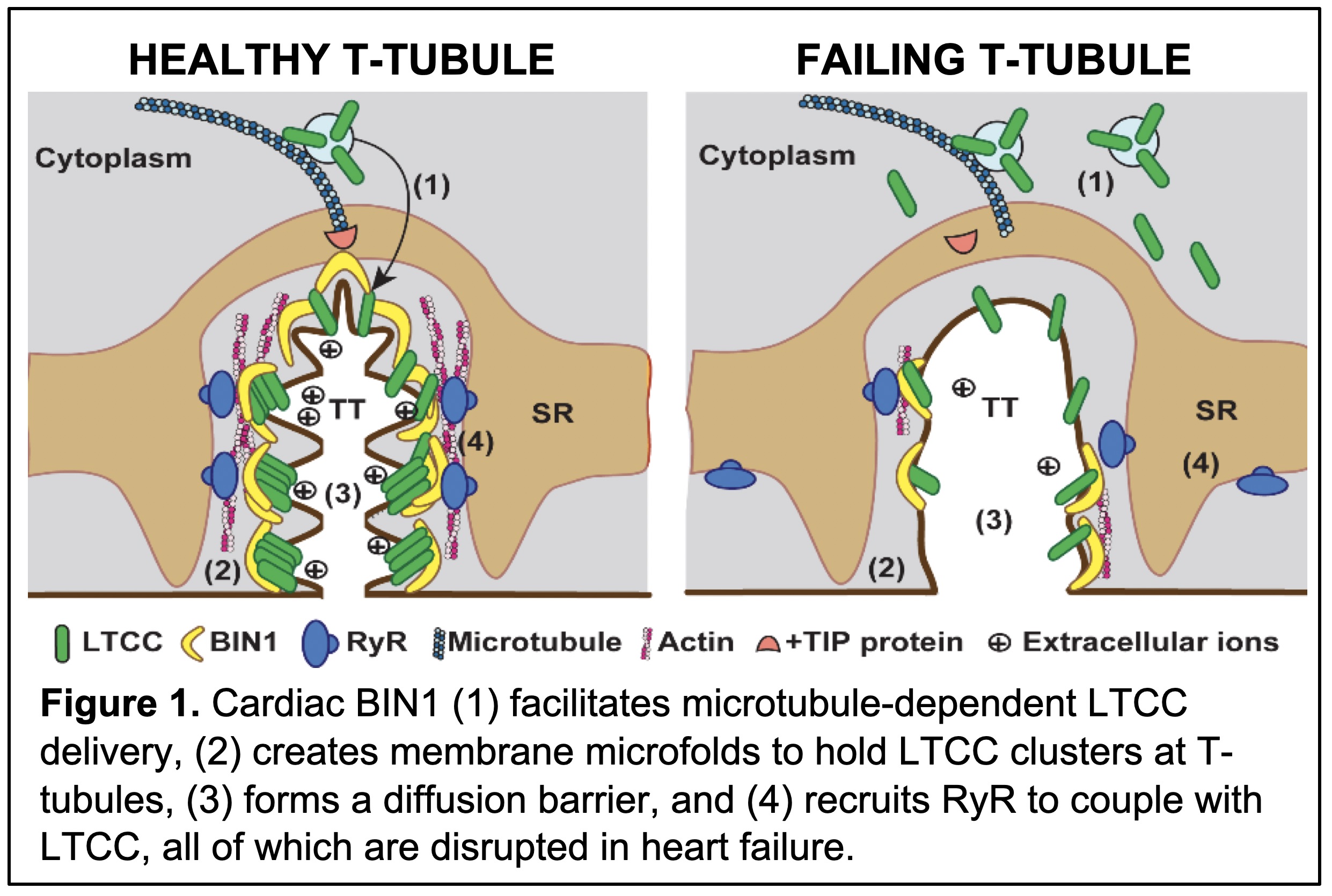 Transverse-tubules (T-tubules) are membrane invaginations specific to striated muscles.
Cardiac T-tubules are enriched with L-type calcium channels (LTCCs), which form dyads
with ryanodine receptors (RyRs) to initiate intracellular calcium signaling critical
for the beat-to-beat heart contraction. We previously identified that a membrane curvature
protein, the cardiac isoform of BIN1 (Bridging Integrator 1) now called cBIN1, folds
the membrane bilayer within T-tubules to control extracellular ion diffusion, protecting
the hearts from arrhythmias (Figure 1). Over the years, we have identified that cBIN1-microdomains
regulate myocardial function by facilitating microtubule-dependent LTCC delivery,
clustering LTCCs and recruiting RyRs for effective dyad formation, and organizing
SERCA2a for effective diastolic calcium removal and myocardial relaxation. In heart
failure, disruption of cBIN1 microdomains causes impaired myocardial function, which
can be normalized by cBin1 gene therapy. We are now further resolving the fundamental cell biology underlying
cBIN1-microdomain formation, regulation, and turnover. The translational goal is to
explore new strategies, via targeting cBIN1-microdomains, to develop novel therapies
effective for patients with heart failure.
Transverse-tubules (T-tubules) are membrane invaginations specific to striated muscles.
Cardiac T-tubules are enriched with L-type calcium channels (LTCCs), which form dyads
with ryanodine receptors (RyRs) to initiate intracellular calcium signaling critical
for the beat-to-beat heart contraction. We previously identified that a membrane curvature
protein, the cardiac isoform of BIN1 (Bridging Integrator 1) now called cBIN1, folds
the membrane bilayer within T-tubules to control extracellular ion diffusion, protecting
the hearts from arrhythmias (Figure 1). Over the years, we have identified that cBIN1-microdomains
regulate myocardial function by facilitating microtubule-dependent LTCC delivery,
clustering LTCCs and recruiting RyRs for effective dyad formation, and organizing
SERCA2a for effective diastolic calcium removal and myocardial relaxation. In heart
failure, disruption of cBIN1 microdomains causes impaired myocardial function, which
can be normalized by cBin1 gene therapy. We are now further resolving the fundamental cell biology underlying
cBIN1-microdomain formation, regulation, and turnover. The translational goal is to
explore new strategies, via targeting cBIN1-microdomains, to develop novel therapies
effective for patients with heart failure.
Microdomain Turnover and cBIN1 Extracellular Vesicles
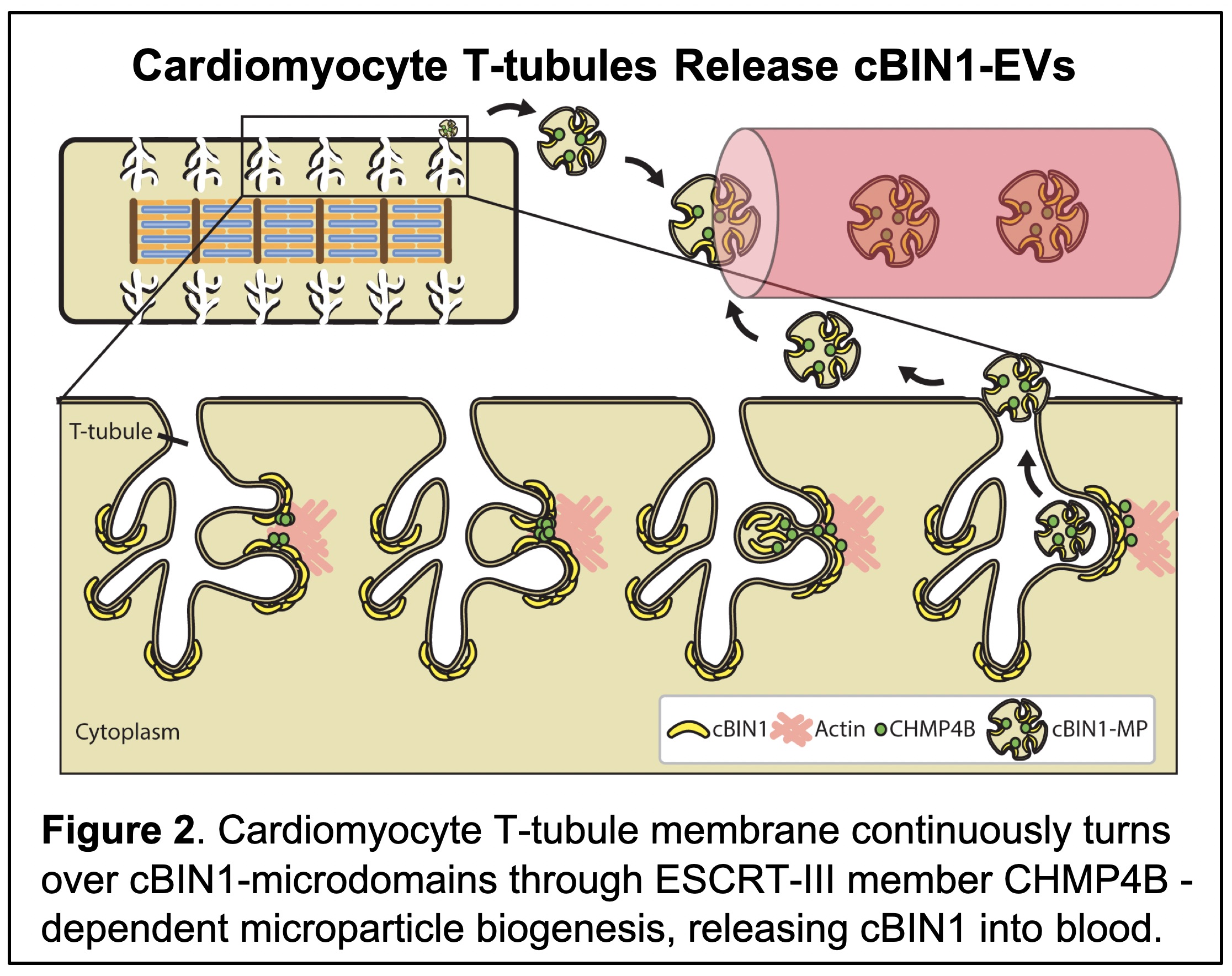 On the other hand, cardiomyocyte T-tubule cBIN1-microdomains turn over through microparticle
release, and blood cBIN1 can aid in diagnosis and prognosis of heart failure. We also
identified that cBIN1-microparticle release involves the ESCRT-III subunit CHMP4B
(Figure 2), which is expressed in the mammalian cardiomyocytes and interacts with
cBIN1. For the first time, the BAR domain containing protein superfamily is found
to serve as an early ESCRT factor to initiate the biogenesis of extracellular vesicles.
These results introduce a new paradigm that cardiomyocyte membrane undergoes dynamic
turnover, releasing membrane microdomains. We are currently further exploring the
release mechanism, functional significance, and therapeutic potential of these cardiac
origin cBIN1 vesicles.
On the other hand, cardiomyocyte T-tubule cBIN1-microdomains turn over through microparticle
release, and blood cBIN1 can aid in diagnosis and prognosis of heart failure. We also
identified that cBIN1-microparticle release involves the ESCRT-III subunit CHMP4B
(Figure 2), which is expressed in the mammalian cardiomyocytes and interacts with
cBIN1. For the first time, the BAR domain containing protein superfamily is found
to serve as an early ESCRT factor to initiate the biogenesis of extracellular vesicles.
These results introduce a new paradigm that cardiomyocyte membrane undergoes dynamic
turnover, releasing membrane microdomains. We are currently further exploring the
release mechanism, functional significance, and therapeutic potential of these cardiac
origin cBIN1 vesicles.
References (Selected Publications)
- Li J, Agvanian S, Zhou K, Shaw RM, Hong T (2020). Exogenous Cardiac Bridging Integrator 1 Benefits Mouse Hearts With Pre-existing Pressure Overload-Induced Heart Failure. Front Physiol, 11, 708.
- Liu Y, Zhou K, Li J, Agvanian S, Caldaruse AM, Shaw S, Hitzeman TC, Shaw RM, Hong T (2020). In Mice Subjected to Chronic Stress, Exogenous cBIN1 Preserves Calcium-Handling Machinery and Cardiac Function. JACC Basic Transl Sci, 5(6), 561-578.
- Nikolova AP, Hitzeman TC, Baum R, Caldaruse AM, Agvanian S, Xie Y, Geft DR, Chang DH, Moriguchi JD, Hage A, Azarbal B, Czer LS, Kittleson MM, Patel JK, Wu AHB, Kobashigawa JA, Hamilton M, Hong T*, Shaw RM* (2018). (* corresponding authors) Association of a Novel Diagnostic Biomarker, the Plasma Cardiac Bridging Integrator 1 Score, With Heart Failure With Preserved Ejection Fraction and Cardiovascular Hospitalization. JAMA Cardiol, 3(12), 1206-1210.
- Xu B, Fu Y, Liu Y, Agvanian S, Wirka RC, Baum R, Zhou K, Shaw RM, Hong T (2017). The ESCRT-III pathway facilitates cardiomyocyte release of cBIN1-containing microparticles. PLoSBiol, 15(8), e2002354.
- Hong T, Shaw RM (2017). Cardiac T-Tubule Microanatomy and Function. Physiol Rev, 97(1), 227-252.
- Fu Y, Shaw SA, Naami R, Vuong CL, Basheer WA, Guo X, Hong T (2016). Isoproterenol Promotes Rapid Ryanodine Receptor Movement to Bridging Integrator 1 (BIN1)-Organized Dyads. Circulation, 133(4), 388-97.
- Hong T, Yang H, Zhang SS, Cho HC, Kalashnikova M, Sun B, Zhang H, Bhargava A, Grabe M, Olgin J, Gorelik J, Marbán E, Jan LY, Shaw RM (2014). Cardiac BIN1 folds T-tubule membrane, controlling ion flux and limiting arrhythmia. Nat Med, 20(6), 624-32.
- Hong T, Cogswell R, James CA, Kang G, Pullinger CR, Malloy MJ, Kane JP, Wojciak J, Calkins H, Scheinman MM, Tseng ZH, Ganz P, De Marco T, Judge DP, Shaw RM (2012). Plasma BIN1 correlates with heart failure and predicts arrhythmia in patients with arrhythmogenic right ventricular cardiomyopathy. Heart Rhythm, 9(6), 961-7.
- Hong T, Smyth JW, Chu KY, Vogan JM, Fong TS, Jensen BC, Fang K, Halushka MK, Russell SD, Colecraft H, Hoopes CW, Ocorr K, Chi NC, Shaw RM (2012). BIN1 is reduced and Cav1.2 trafficking is impaired in human failing cardiomyocytes. Heart Rhythm, 9(5), 812-20.
- Hong T, Smyth JW, Gao D, Chu KY, Vogan JM, Fong TS, Jensen BC, Colecraft HM, Shaw RM (2010). BIN1 localizes the L-type calcium channel to cardiac T-tubules. PLoS Biol, 8(2), e1000312.