Gabrielle Kardon
Professor of Human Genetics and Adjunct Professor of Nutrition and Integrative Physiology
Musculoskeletal Development, Regeneration

Molecular Biology Program
Education
B.S. Yale University
Ph.D. Duke University
Research
How does muscle develop, regenerate, maintain, age, and evolve? These are the questions that drive our research. We focus on muscle stem cells because they are the source of all muscle. We focus on the muscle connective tissue because it provides the niche for muscle stem cells and is critical for muscle form and function. We study how interactions between muscle stem cells and the connective tissue orchestrate development of limb muscles and the diaphragm, regulate muscle regeneration after injury and viral infection, are the source of birth defects and fibrosis, and shape evolution of the musculoskeletal system.
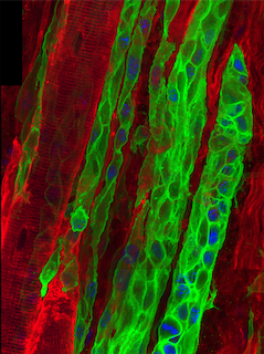
Muscle Stem Cells in Development, Homeostasis, and Regeneration
Muscle development, maintenance, and regeneration take place throughout vertebrate life. Muscle stem cells are critical for all of these processes. Our research has concentrated on the developmental origin of muscle stem cells, their function, and the extrinsic signals that regulate them. We have found that all muscle stem cells in the limb arise in the embryo from the somites, but there are related, although distinct populations of stem cells in the embryo, fetus, and adult (Kardon et al. 2002, Schienda et al. 2006, Hutcheson et al. 2009, Murphy et al. 2011). We also have established that muscle stem cells are critical for muscle development (Hutcheson et al. 2009) and adult muscle stem cells (known as satellite cells) are necessary and sufficient for muscle regeneration (Murphy et al. 2011). Adult muscle stem cells also contribute to muscle homeostasis, although their requirement is still not clear (Keefe et al. 2015). Most recently, we have reconstructed muscle regeneration in three dimensions (Collins, Shapiro et al. 2024). A surprising finding is that the centralized myonuclei are an enduring feature of regenerated myofibers, and we are currently testing whether these myofibers are biomechanically compromised.
Muscle damage and regeneration following alphavirus infection
Ross River (RRV) and Chikungunya (CHIKV) alphaviruses are emerging mosquito-borne pathogens that target the musculoskeletal system and a major health concern as they cause explosive outbreaks in humans. A major unknown in our understanding of RRV and CHIKV disease is how viral infection causes acute and chronic myalgia and myopathy, an important consequence of viral infection found in 90% of patients. In collaboration with Deborah Lenschow’s lab (Washington University, St. Louis) and with initial funding from a Pew Innovation Award, we are determining what functional, structural, cellular, and molecular changes in muscle result from RRV and CHIKV infection and testing whether infection of myogenic cells drives systemic acute and chronic disease.
Diaphragm: Development, CDH, and Evolution
The diaphragm is functionally the most important skeletal muscle in mammals, as it is vital for respiration and serves as a barrier between the thoracic and abdominal cavities. Strikingly, defects in diaphragm development are the cause congenital diaphragmatic hernias (CDHs), a common birth defect (1:300 births) that results in severe morbidity and 50% mortality. Given the diaphragm’s functional importance and the frequency and severity of CDH, we focus on understanding the embryonic origins, cell-cell interactions, and genetic mechanisms underlying diaphragm development normally and during CDH. Using mouse genetics, we definitively established that the pleuroperitoneal folds (PPFs), transient embryonic structures, and the muscle connective tissue fibroblasts derived from them critically regulate development of the diaphragm muscle (Merrell et al. 2015). Furthermore, we showed that mutations in Gata4 in PPF fibroblasts cause CDH. More recently, we have used whole-genome sequencing to comprehensively reconstruct the genetic architecture of 4 CDH individuals and demonstrate that the etiology of CDH is heterogeneous and multifactorial (Bogenschutz et al. 2020). Also, we have developed a novel culture system to efficiently test the functional significance of candidate CDH genes (Bogenschutz et al, 2020). Finally, using conditional mutagenesis in mice, we have established that PPF expression of HGF is essential for recruitment and expansion of muscle in the developing diaphragm (Sefton et al. 2022). Currently, we are further exploring the important molecular interactions between PPF fibroblasts and muscle cells during normal diaphragm development and CDH.
Although the diaphragm is a functionally essential skeletal muscle in humans, the muscularized diaphragm is an evolutionary novelty that arose in and uniquely defines mammals. What genetic and developmental innovations led to this major mammalian innovation? The question of how morphologic novelties arise is a major question in evolutionary biology. To discover what developmental innovations led to the evolution of the muscularized diaphragm, we are comparing the development of birds (chick), which represent an ancestral, non-muscularized state, with mice.
References
- Collins BC*, Shapiro JB, Scheib MM, Musci RV, Verma M, Kardon G. (2024). Three-dimensional imaging studies in mice identify cellular dynamics of muscle regeneration. Developmental Cell Mar 29:S1534-5807(24)00184-9.
- Burns NG* and Kardon G. (2023). The role of genes and environment in the etiology of congenital diaphragmatic hernias. Curr Top Dev Biol 152:115-138.
- Burns NG* and Kardon G. (2023). The roles of genes and environment in the etiology of congenital diaphragmic hernias. Current Topics in Developmental Biology 152: 115-138.
- Sefton EM*, Gallardo M, Tobin CE, Colasanto MP*, Collins BC*, Merrell AM*, Kardon G. (2022). Fibroblast-derived HGF integrates muscle and nerve development during morphogenesis of the mammalian diaphragm. eLife. Sep 26;11:e74592.
- Bogenschutz EL*, Fox ZD, Farrell A, Wynn J, Moore B, Yu L, Aspelund G, Marth G, Yandell M, Shen Y, Chung WK, Kardon G. (2020). Deep whole-genome sequencing of multiple proband tissues and parental blood reveals the complex genetic etiology of congenital diaphragmatic hernias. HGG Advances Oct 22;1(1):100008.
- Bogenschutz EL*, Sefton EM*, Kardon G. (2020). Cell culture system to assay candidate genes and molecular pathways implicated in congenital diaphragmatic hernias. Dev Biol Nov 1;467(1-2):30-38.
- Sefton EM* and Kardon G. 2019. Connecting muscle development, birth defects, and evolution: An essential role for muscle connective tissue. Current Topics in Developmental Biology 132:137-176.
- Sefton EM*, Gallardo M, Kardon G. (2018). Developmental origin and morphogenesis of the diaphragm, an essential mammalian muscle. Dev. Biol. pii: S0012-1606(18)30146-5.
- Kardon G, Ackerman AG, McCulley DJ, Shen Y, Wynn J, Shang L, Bogenschutz E*, Sun X, Chung WK. (2017). Congenital diaphragmatic hernias: from genes to mechanisms to therapies. Dis Model Mech 1;9 (11): 955-970.
- Colasanto MP*, Eyal S, Mohassel P, Bamshad M, Bonnemman C, Zelzer E, Moon AM, Kardon G. (2016). Development of a subset of forelimb muscles and their attachments sites requires the ulnar-mammary syndrome gene Tbx3. Disease Models and Mechanisms 9(11): 1257-1269
- Domyan ET, Kronenberg Z, Infante CR, Vickrey AI, Stringham SA, Bruders R, Guernsey MW, Park S, Payne J, Beckstead RB, Kardon G, Menke, Yandell M, Shapiro (2016). Molecular shifts in limb identity underlie development of feathered feet in two domestic avian species. eLife, 5.
- Keefe AC*, Lawson JA, Flygare SD, Fox ZD, Colasanto MP*, Mathew SJ*, Yandell M, Kardon G (2015). Muscle stem cells contribute to myofibres in sedentary adult mice. Nat Commun, 6, 7087.
- Pawlikowski B, Pulliam C, Betta ND, Kardon G, Olwin (2015). Pervasive satellite cell contribution to uninjured adult muscle fibers. Skelet Muscle, 5, 42.
- Murphy MM*, Keefe AC*, Lawson JA, Flygare SD, Yandell M, Kardon G (2014). Transiently active Wnt/beta-catenin signaling is not required but must be silenced for stem cell function during muscle regeneration. Stem Cell Reports, 3(3), 475-88.
- Rohatgi A, Corbo JC, Monte K, Higgs S, Vanlandingham DL, Kardon G, Lenschow DJ (2014). Infection of myofibers contributes to increased pathogenicity during infection with an epidemic strain of chikungunya virus. J Virol, 88(5), 2414-25.
- Merrell AJ*, Kardon G (2013). Development of the diaphragm -- a skeletal muscle essential for mammalian respiration. FEBS J, 280(17), 4026-35.
- Wan Y*, Lewis AK, Colasanto M*, van Langeveld M, Kardon G, Hansen C (2012). A practical workflow for making anatomical atlases for biological research. IEEE Computer Graphics and Applications, 32(5), 70-80.
- Murphy MM*, Lawson JA, Mathew SJ*, Hutcheson DA*, Kardon G (2011). Satellite cells, connective tissue fibroblasts and their interactions are crucial for muscle regeneration. Development, 138(17), 3625-37.
- Mathew SJ*, Hansen JM, Merrell AJ*, Murphy MM*, Lawson JA, Hutcheson DA*, Hansen MS*, Angus-Hill M, Kardon G (2011). Connective tissue fibroblasts and Tcf4 regulate myogenesis. Development, 138(2), 371-84.
- Murphy M*, Kardon G (2011). Origin of vertebrate limb muscle: the role of progenitor and myoblast populations. Curr Top Dev Biol, 96, 1-32.
- Hutcheson DA*, Kardon G (2009). Genetic manipulations reveal dynamic cell and gene functions: Cre-ating a new view of myogenesis. Cell Cycle, 8(22), 3675-8.
- Hutcheson DA*, Zhao J, Merrell A*, Haldar M, Kardon G (2009). Embryonic and fetal limb myogenic cells are derived from developmentally distinct progenitors and have different requirements for beta-catenin. Genes Dev, 23(8), 997-1013.
- Schienda J, Engleka KA, Jun S, Hansen MS*, Epstein JA, Tabin CJ, Kunkel LM, Kardon G (2006). Somitic origin of limb muscle satellite and side population cells. Proc Natl Acad Sci U S A, 103(4), 945-50.
- Kardon G, Harfe BD, Tabin CJ (2003). A Tcf4-positive mesodermal population provides a prepattern for vertebrate limb muscle patterning. Dev Cell, 5(6), 937-44.
- Kardon G, Campbell JK, Tabin CJ (2002). Local extrinsic signals determine muscle and endothelial cell fate and patterning in the vertebrate limb. Dev Cell, 3(4), 533-45.